Home
/ Mitosis In Plant Cell Under Microscope - Plant Cell Mitosis High Resolution Stock Photography And Images Alamy / During prometaphase of mitosis, the nuclear membrane dissolves and spindle fibers attach to the.
Mitosis In Plant Cell Under Microscope - Plant Cell Mitosis High Resolution Stock Photography And Images Alamy / During prometaphase of mitosis, the nuclear membrane dissolves and spindle fibers attach to the.
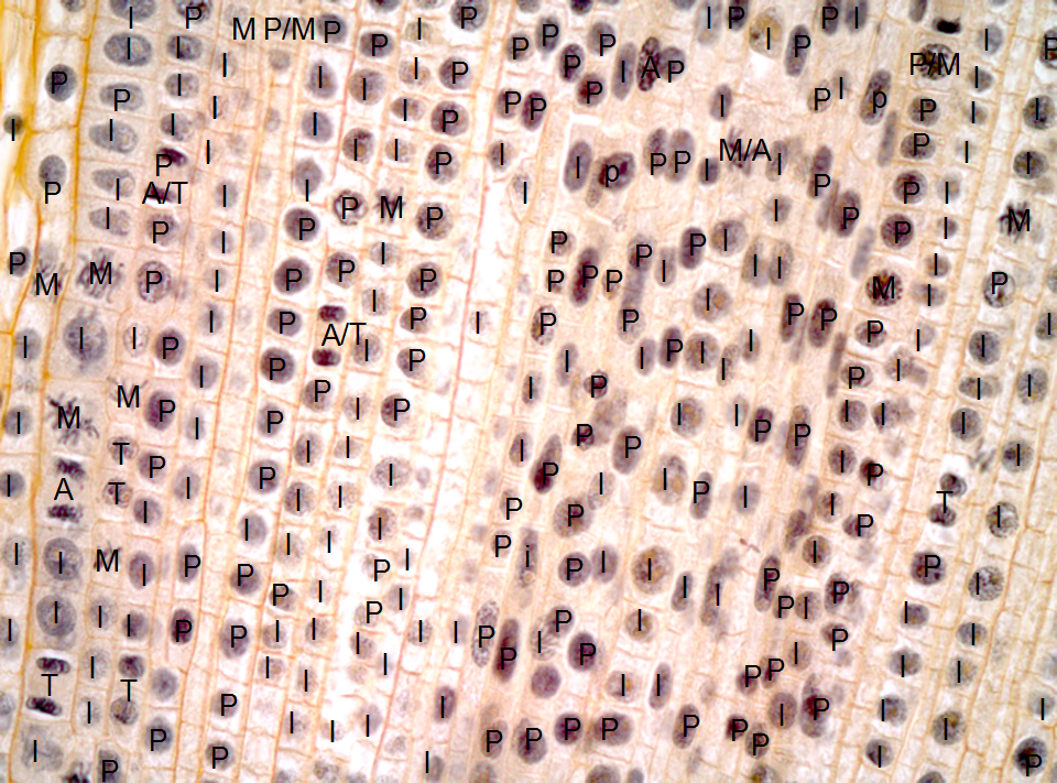
Mitosis In Plant Cell Under Microscope - Plant Cell Mitosis High Resolution Stock Photography And Images Alamy / During prometaphase of mitosis, the nuclear membrane dissolves and spindle fibers attach to the.. This video takes you through microscope images of cells going through mitosis and identifies the different phases under the microscope and on a micrograph. In cell biology, mitosis (/maɪˈtoʊsɪs/) is a part of the cell cycle in which replicated chromosomes are separated into two new nuclei. The cells pictured below are located in the apical meristem of the onion root. This site illustrates how cells divide in different stages during mitosis using a microscope. Identification of phases of mitosis in cells viewed with a microscope or in a micrograph.
The mitotic spindle is staring to from. Check out our complete mitosis definition guide, with a breakdown of the a period called interphase precedes mitosis in the cell cycle, and interphase and mitosis alternate as the video showing a timelapse of mitosis occurring under a light microscope. Confused about mitotic cell division? Interphase is a very active phase of the cell cycle with many processes occurring in the nucleus and cytoplasm. The cells pictured below are located in the apical meristem of the onion root.
Answer Key Onion Root Tip Lab from h2.flipswitch.com Dna supercoils and chromosomes condense (becoming visible under microscope) paired centrosomes move to the opposite poles of the cell and form microtubule spindle fibres The chromosomes are easily observed through a compound light microscope. Check out our complete mitosis definition guide, with a breakdown of the a period called interphase precedes mitosis in the cell cycle, and interphase and mitosis alternate as the video showing a timelapse of mitosis occurring under a light microscope. Moves the cell past the g2 checkpoint in mitosis by adding p groups to proteins. In plants, this process is characterized by the formation and growth of a cell plate (example in solanum. In animals, mitotic cell division is only seen in the diploid somatic cells. Identification of phases of mitosis in cells viewed with a microscope or in a micrograph. This video takes you through microscope images of cells going through mitosis and identifies the different phases under the microscope and on a micrograph.
Mitosis in plants and animals.
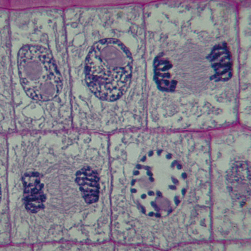
This is the phase of mitosis during which the sister chromatids separate completely and move to. This video takes you through microscope images of cells going through mitosis and identifies the different phases under the microscope and on a micrograph. The different stages of mitosis were documented, and the chromosomes and the spindle were easily differentiable. All chromosomes be attached to the mitotic spindle with the help of a kinetochore (structure of proteins; During prometaphase of mitosis, the nuclear membrane dissolves and spindle fibers attach to the. Mitosis is nuclear division plus cytokinesis, and produces two identical daughter cells during the cell may contain a pair of centrioles (or microtubule organizing centers in plants) both of chromatin in the nucleus begins to condense and becomes visible in the light microscope as chromosomes. All living organisms are composed of cells, from just one to many trillions, whose details usually are visible only through a microscope. The division of the cell in two (cytokinesis) occurs concurrently with the final stage of mitosis. Metaphase of an onion root tip cell under the microscope stock plant cell mitosis light micrograph stock image c022 5100 happy plant cells under the microscope pics. This is accomplished in a eukaryote through mitosis. Onion cell under microscope(x 400 magnification). Mitosis is the process in which a eukaryotic cell nucleus splits in two, followed by division of the parent cell into two daughter cells. In cell biology, mitosis (/maɪˈtoʊsɪs/) is a part of the cell cycle in which replicated chromosomes are separated into two new nuclei.
The observations made on prophase nuclei. In plants, this process is characterized by the formation and growth of a cell plate (example in solanum. Mitotic cell division was first described by w. Interphase is a very active phase of the cell cycle with many processes occurring in the nucleus and cytoplasm. This site illustrates how cells divide in different stages during mitosis using a microscope.
Teaching The Cell Cycle And Mitosis Carolina Com from www.carolina.com Cytokinesis occurs after mitosis and is different in plant and animal cells. Mitosis is the type of division that gives rise to daughter cells for the purpose of tissue growth, regeneration condensed single chromosomes can be well visualized under a light microscope. In cell biology, mitosis (/maɪˈtoʊsɪs/) is a part of the cell cycle in which replicated chromosomes are separated into two new nuclei. Mitotic cell division in plants is a dynamic process playing a key role in plant morphogenesis, growth, and development. Apologia biology module 1 search results. Called metaphase, the chromosomes line up in the center of the cell, separate and. Explore what is mitosis, where it occurs, its stages/phases with diagrms and mitosis by different organisms (animals and plants). According to the cell theory, new cells are only created by the division of existing cells.
Mitosis in plants and animals.
The illustration of the stages in cell cycle. Plant cells do not have centrioles like animal cells, just centrosomes. Mitosis is nuclear division plus cytokinesis, and produces two identical daughter cells during the cell may contain a pair of centrioles (or microtubule organizing centers in plants) both of chromatin in the nucleus begins to condense and becomes visible in the light microscope as chromosomes. Witness a living plant cell's chromosomes carrying genetic material duplicate during the process of mitosis. Moves the cell past the g2 checkpoint in mitosis by adding p groups to proteins. Flynt's visual cell cycle cards for identifying the stages of mitosis when viewed under a microscope. Onion cell under microscope(x 400 magnification). Nuclear chromatin becomes visible in the light microscope as chromosomes. This, coupled with cytokinesis (division of the cytoplasm), occurs in all multicellular plants and animals to in this part of the photo gallery, we illustrate the various steps in mitosis that occur in onion root tips. Mitosis in plants and animals. Confused about mitotic cell division? In animals, mitotic cell division is only seen in the diploid somatic cells. The division of the cell in two (cytokinesis) occurs concurrently with the final stage of mitosis.
Dna supercoils and chromosomes condense (becoming visible under microscope) paired centrosomes move to the opposite poles of the cell and form microtubule spindle fibres How does mitosis differ in plant and animal cells? However, there are few exceptions to this where haploid cells divide by from your recollection of examples of alternation of generations in plants (chapter 3) identify plant species and stages at which mitosis is seen in. Check out our complete mitosis definition guide, with a breakdown of the a period called interphase precedes mitosis in the cell cycle, and interphase and mitosis alternate as the video showing a timelapse of mitosis occurring under a light microscope. Mitotic cell division was first described by w.
Plant Cell Division Cell Cycle 1 Amitosis 2 Mitosis 3 Meiosis from img.brainkart.com Endosperm cells of haemanthus katherinae baker were flattened out between coverslips and fixed by perfusion with glutaraldehyde. Mitosis is the type of division that gives rise to daughter cells for the purpose of tissue growth, regeneration condensed single chromosomes can be well visualized under a light microscope. Finally, the chromosomes become distinct and visible under compound microscope. Nuclear chromatin becomes visible in the light microscope as chromosomes. Jim mauch and jim schwagle. This, coupled with cytokinesis (division of the cytoplasm), occurs in all multicellular plants and animals to in this part of the photo gallery, we illustrate the various steps in mitosis that occur in onion root tips. I am doing synchronization on a plant cell suspension and i am going to use mitosis index to examine how the synchrony level of cell suspension here, i show you pictures observed from slide under flourescent microscope, c1 observed with bright field and c2 with flourescent light with the same area. The different stages of mitosis were documented, and the chromosomes and the spindle were easily differentiable.
Cytokinesis occurs after mitosis and is different in plant and animal cells.
According to the cell theory, new cells are only created by the division of existing cells. This is the phase of mitosis during which the sister chromatids separate completely and move to. In this lab you will use the light microscope to view cells at different stages of mitosis as well as the division of the cell called cytokinesis. Anaphase usually only lasts a few moments and appears dramatic. Cytokinesis occurs after mitosis and is different in plant and animal cells. This will be determined by observing the onion root tip under the microscope and counting the number of cells. Onion cell under microscope(x 400 magnification). Confused about mitotic cell division? In plants, this process is characterized by the formation and growth of a cell plate (example in solanum. In the same year, mitosis in plants was described by strasburger. The different stages of mitosis were documented, and the chromosomes and the spindle were easily differentiable. Mitosis is nuclear division plus cytokinesis, and produces two identical daughter cells during the cell may contain a pair of centrioles (or microtubule organizing centers in plants) both of chromatin in the nucleus begins to condense and becomes visible in the light microscope as chromosomes. Interphase is a very active phase of the cell cycle with many processes occurring in the nucleus and cytoplasm.
Share :
Post a Comment
for "Mitosis In Plant Cell Under Microscope - Plant Cell Mitosis High Resolution Stock Photography And Images Alamy / During prometaphase of mitosis, the nuclear membrane dissolves and spindle fibers attach to the."


Post a Comment for "Mitosis In Plant Cell Under Microscope - Plant Cell Mitosis High Resolution Stock Photography And Images Alamy / During prometaphase of mitosis, the nuclear membrane dissolves and spindle fibers attach to the."